Tapejara: abdominal bones

These are experimental. There are not many museum casts out there with gastralia and pre-pubis. Not many pre-pubis bones are preserved, either. I used as a source the MN 6588-V tapejarid, scaled for Tapejara. It shows a pair of pre-pubis bones. They are flat, so I can only speculate about their shape, but since they articulate with the pubis, and gastralia bones, which articulate with the ribs to make a rounded belly, I believe they are concave on the ventral side. That’s also what reconstructions of other pterosaurs suggest. I am not sure yet if I will be able to use them, since there is a lot of speculation in that part of Tapejara’s body (unknown number of dorsals, sternal ribs, etc.)

Here is the pre-pubis I cut out of a 2mm sheet of foam, based on the MN 6588-V specimen.

2013-07-24 01.18.16

I made gastralia from pieces of foam like this.

2013-08-18 09.52.51

They are probably not thin enough. But hey, maybe this Tapejara had a stiff belly 🙂

2013-08-22 17.55.14

2013-08-22 17.55.25

1 Comment

Filed under Pterosaur #7: Tapejara

Tapejara: pelvic girdle

I already had made the pelvic bones, but the sternum complex was missing. I didn’t find any realiable source with a perfect sternum, so I used as a source the same azhdarchoid sternum that I used in Tupuxuara, and adjusted it so it would fit the Tapejara pelvis (from SMNK PAL 1137). They are 5 sacral vertebrae, and two dorsal ones with lateral processes fused to the preacetabular processes of the ilium.

I cut it out in one piece from a sheet of 2mm foam, and added the stacked vertebral bodies sculpted out of thicker (yellow) foam. Here are two views of the sacrum after treating with fire.

sacrum 2 sacrum 1

Four views of the finished sacrum.

lateral ventral posterior anterior

Next step: assemble the pectoral girdle. I now have all the parts.

parts

I wish I had smaller pins.

testing dorsal dorsal w pelvis testing lateral anterior with pelvis posterior with pelvis

I placed a rubber tube through the vertebrae acting as a spinal cord. It will be used to attach the pelvic girdle to the dorsal spine and to attach the tail on the other side.

ventral w pelvis

I now can test all the connections. Here is the Tapejara dressed in its pelvic girdle.pelvis and sacrum

A dorsal view.

dorsal assembly

And a ventral one. It’s almost finished. I’m already prototyping the pre-pubis and gastralia, but before I assemble the rest I will cover what I have so far with epoxi resin (I can’t use pins anymore).

ventral assembly lateral assembly

1 Comment

Filed under Pterosaur #7: Tapejara

Tapejara: pectoral girdle

The pectoral girdle consists of scapula, coracoid, sternum, dorsal vertebrae and ribs that grow from the vertebrae and from the sternum. I’m still having trouble figuring out how to assemble the scapula and coracoid. I read some articles and I haven’t discovered yet what most paleontologists think is the best layout. They seem to disagree in many aspects. Without twisting the bones quite a bit or setting the scapula to articulate with the first two dorsals I can’t obtain a low position. At best, the glenoid will stay somewhere in the middle of the chest. For now, I just pinned the proximal end of the scapula to the third dorsa, and the distal end of the coracoid to the sternum, so I could work on the rest of the pectoral girdle. But the correct way to connect scapula and coracoid is still an issue I haven’t resolved. Any help is welcome.

The ribs. I found some random ribs in fossils, but I preferred to use Wellnhofer’s reconstruction of Pteranodon as a reference to start with. I scaled them for Tapejara and made these sketches.

rib cut out

I cut out the two first pairs.

rib cut out 2

I made them hollow of course. This is a first prototype attached to the first dorsal vertebra.

rib testing

I quickly made some (generic) sternal ribs and connected their ends to the sternum and larger ribs to test the shape of the rib cage.

girdle testing girdle testing 3

Time to make more ribs.

more cut outs 2 first rib set

testing

The sternal ribs are thin but they have some lateral very thin bone pieces projecting laterally. It looks like some kind of ossified cartilage. How many ribs should be connected to the sternum? Some say seven lateral pairs, many reconstructions show four or five. The azhdarchoid MN-6588-V seems to have seven, and probably two more pairs behind. I chose five.

sternal ribs 2

Now the challenging attempt to assemble the pectoral girdle.

pectoral assembly parts 2

Somehow I fitted the scapula and coracoid in a way that revealed a glenoid and didn’t seem weird. The proximal end of the scapula is pinned to the third dorsal vertebra. If I project it a bit more laterally, it can move back more, but the sternum-coracoid articulation will also change.

ventral body 2

So now it looks like this.

ventral body lateral body

1 Comment

Filed under Pterosaur #7: Tapejara

Tapejara: shoulders and chest

I had two sources for the scapula and coracoid, and three for the sternum. For the scapula and coracoid I initially used SMNK PAL 1137 but then received better photos from IMCF 1061. The scapulae looked alike, but the proximal end of the coracoid was very different, it was also much longer. I don’t know if any part is missing or if it is just a result of SMNK PAL 1137 representing a very young specimen. Anyway, it is still very hard to identify and compare certain details when one photo is a bad black and white one with only two views. I decided to make the scapula and coracoid from IMCF 1061, and the sternum from SMNK PAL 1137 scaled by SMNK PAL 3985.

After estimating widths and depths from the photos, these are the flat parts I came up with. The yellow foam was used for the ends, which I had to sculpt.

cut outs scapulocoracoid

The parts after cutting out.

parts

Assembly of the scapula.

scapula assembly

Assembly of the coracoid.

coracoid assembly

Partial assembly of scapulae and coracoids.scap cora assemblies

This is the distal end of the scapula.

distal scap

And here is a set of coracoid and scapula after treatment with fire:

burned scap cora

Since there were many photos, I was able to compare several angles, and see where it was curved, thick, thin, and other details. This is another side view of the scapula.

side burned scap

The sternum is quite flat, so I initially made a shape slightly larger than the photo, so I could then give it a curved aspect.

stern cut out

But the keel I had to make separately.

keel

Here is a picture of the sternum with the keel attached shortly after treating with fire.

anterior sternum keel

And here is a side view after applying modelling paste and coffee stains.

lateral sternum

Compare it with the humerus.

sternum humerus

Now I have a big problem. How to assemble the pectoral girdle. I still didn’t figure out exactly which ends of the scapula and coracoid connect to form the scapulocoracoid. I might still have to twist the bone a bit since I may not have captured all the three-dimensional details from the photos. I spent a couple of hours trying different positions and gave up. Tomorrow I will try to assemble the pelvic girdle with ribs, and it might be easier. Here are the final photos.

dorsal sternum ventral sternum anterior sternum lateral scapula

1 Comment

Filed under Pterosaur #7: Tapejara

Tapejara carpals and pteroid

I used as sources only the black and white pictures from SMNK PAL 1137 published by Eck et al (2011) which shows four views of distal and proximal syncarpals, but only two views of the medial carpal (which I assume are perpendicular views.) I’ve had many problems with the scales in this article, but I tested the proximal and distal syncarpals with the bones that articulate with them (wing metacarpal, radius and ulna). I am not so sure about the medial carpal, but it seems to fit in a gap on the anterior part of the assembly, so the pteroid fits between the medial carpal and the proximal one.

I made the pteroid hollow, of course. I used IMCF 1061 as a source.

2013-08-13 13.55.17

Here are the wrist bones. I might make some adjustments. I will publish more pictures later showing the connections.

2013-08-13 14.17.24

1 Comment

Filed under Pterosaur #7: Tapejara

Tapejara’s hands and feet

It was challenging to make such small bones. The metacarpals were the thinnest ones and the phalanges of the feet the smallest. I obviously didn’t make them hollow since they are thinner than my 2mm thick sheet of foam, but I did try to. I made hollow fingers, but later reduced their width by pressing and by using fire. That made them stronger (although the result was a bit thicker than I expected). Since they are small and very fragile, I can’t pin them together (I would need some micropins for that), so I already covered them with epoxi resin. Here are the pictures.

This is a metatarsi from one of the feet, before treating with fire and resin.

Metatarsal

Here I try to fit the bones in place to make adjustments. The images from SMNK PAL 1137 are very bad but I used it to get the proportions of the metatarsi and the first toe phalanx and toenail. I initially then used as sources individual photos of Tapejara foot bones, but they are usually in unprepared blocks and not always entirely visible, and sometimes damaged. So my measurements didn’t match. I also looked at several well-preserved photos of different species (like SMNK PAL 3830) but then adapted a generic layout from Wellnhofer’s handbook for Pteranodon, scaled for Tapejara. It seems that the smalled phalanges are a bit smaller in Tapejara, and I tried to make them a bit smaller. Here are the bones of the feet after attempting an assembly.

assembly

And these are the hand fingers (I made the phalanges hollow).

finger assembly

I pinned the fingers to the wing metacarpal to try it out.

hand testing 2

hand testing

Then I pinned all these little bones on a surface to apply the epoxi resin.

resin 4

I feel like leaving it this way 🙂 A Tapejara bone graveyard under an unknown Azhdarchoid skull.

resin 6

Here are all the shiny parts after the resin dried.

shiny

These are the toe bones. Each square measures 5mm (remember this Tapejara was scaled up 25%).

toe set

And the bones in place (I will make more adjustments when I connect the bones together with silicone rubber).

toe assembly

Get these rocks and assemble a pair of hands?

finger set 2

finger and metacarpal 2

1 Comment

Filed under Pterosaur #7: Tapejara

Tapejara pelvis and legs

I’m still don’t have all the information for the pelvis, but I decided to start doing it anyway. My only source are the images of SMNK PAL 1137 from the article by Eck et al, which are in black and white, low resolution and have at most two views of each bone. I will have to use what I know about other pterosaurs to make it three-dimensional.

I started cutting out the parts in scale (the scale is another big problem – I based my choice on information from paleontologists since the scales in the article are not reliable).

2013-07-23 22.03.38

Then I  folded and tried to shape each bone trying to guess the depth of its three-dimensionality from the shadows. I also used as references all I learned about pterosaur pelves so far, but I never made one from the individual bones before.

2013-07-24 01.20.01

This is a prototype. The pelvic bones are actually a set of four bones on each side forming the pterosaur’s hips. It’s supposed to look something like this. The long one on the left is the ilium (the long part of the ilium is actually called the preacetabular process). On the right, above is the postacetabular process. The depression near the right side of the ilium, which forms a circular shape if you consider the other two bones below is the acetabulum. That’s where the femur will articulate. At the lower left is the pubis and at lower right, the ischium. The point where the ilium, pubis and ischium fuse together is the acetabulum.

2013-07-24 02.40.57

Here I added some more detail, did some fire shaping and coffee-staining. It’s still a prototype for testing (I might change it or throw it away after I use it). You can see better now the three-dimensional aspect of the ilium, showing the acetabular depression, and the medial side of the postacetabular process.

2013-07-26 19.46.02

The process I used to make the femora and tibiae is the same I used for the other long bones (humeri, ulnae, etc) so I won’t detail them here. This is a picture of the strips of foam I used to make the bones of the legs. One of the femora is already with its halves glued in place.

2013-07-24 01.17.39

The tibia is actually a set of four bones called the tibiotarsus. It contains the tibia itself, the fibula (a very thin bone fused to one of the sides of the tibia, starting at the proximal end and not reaching the distal end), the calcaneum and astragalus which are actually part of the feet (they are the proximal tarsi which function as hinges for the feet). The picture below shows the tibia-fibula and the calcaneum-astragalus parts.

2013-07-26 00.30.38

Now we can try assembling the whole set, so see how it looks. In the upper part of the image below is an unassembled pelvis, a femur and a tibiotarsus, and at the bottom an assembled pelvis with an articulated femur and tibiotarsus.

2013-07-26 19.55.46

Here I tried to fit them in place with my fingers.

2013-07-26 22.36.41

But it’s easier with pins. The image below shows a tibiotarsus, femur, and an assembled pelvis.

2013-07-26 23.45.38

There is still a lot of work to do on the pelvis, and I should return to it next week when I will try to connect them to the sacrum, and finish the pelvic girdle.

1 Comment

Filed under Pterosaur #7: Tapejara

Tapejara wings

The wing is a giant finger, and fingers are connected to the radio/ulna pair by several small bones called carpals and metacarpals. I’ll leave the carpals for another opportunity and concentrate on the metacarpal and four phalanges of the wing.

The wing metacarpal is a big bone, about the size of the radius and ulna and even wider on one end. The wider end articulates with the radio-ulna pair through the carpal bones, and the other is a hinge for the first phalange of the wing – the longest bone in the Tapejara body.

I used SMNK PAL 1137 as a source, but had to rely on Tupuxuara for some details. They are similar. I got the length dimensions from Brian Andres’s database (scaled 25%). I used the same process I employed on the other long bones. Cut two halves, shape, glue together, fix with fire, add ends in thicker foam and reshape.

IMG_5371 IMG_5376 IMG_5381 2013-07-24 18.00.09

After the metacarpal comes the long first phalanx. This one I could rely on a picture I have from IMCF 1061 (which was perfect, since the quality of the pictures in SMNK PAL 1137 is very bad).

This bone has a flattened shaft, so it has greater resistance to anteroposterior forces than to dorsoventral ones.

IMG_5350

You can see I added a thin strip of plastic to the middle of the bone. Does that increase resistance? I measured it. A strip of 2mm foam 20 cm long  and 7mm wide attached on one end (1cm for attachment) can support 7 grams on the other end before folding (forcing the weak narrower side). Treating it with fire made it resist more, but it cracked with 9 grams. The phalanx has two of these strips, and is a bit hollow (I glue them on the edges, slightly curved). I made a prototype which supported 20 grams before cracking. Adding the strip won’t keep it from bending, but will avoid a destructive bend in one place or cracking. I expect that resistance to increase when I add the acrylic resin, and even more after the epoxy resin coating. Anyway, the maximum of weight the phalange will have to bear (considering only support on the proximal end) is 7 grams (considering the other three phalanges already coated with resin and epoxy and connected with silicone rubber). They currently weigh less than 5 grams (no epoxy coating yet) and they still have some water from the acrylic resin to evaporate.

IMG_5351

Here are the finished bones of the first phalanx compared to an unfinished humerus and the neurocranium.

IMG_5359

Some closeups and other angles.

IMG_5363 IMG_5361

Testing the articulation with the metacarpal.

IMG_5379 IMG_5378

The articulation with the radius-ulna pair is still not possible because there are yet no carpals.

2013-07-25 16.48.45

Next step: the other three phalanges. In fact I already had them all cut out and the haves attached before I did metacarpals and radius-ulna pairs. All these unfinished bones fit nicely on top my computer.

2013-07-24 11.38.47

So I tested the wings before I made these bones.

IMG_5396

And here is the final result. I used IMCF 1061 as sources for phalanges 2 and 3, and Tupuxuara for the last phalanx. I also used measurements from Brian Andres (scaled 25% as usual, since this is a larger specimen).

2013-07-25 16.48.35

Here is a detail of the last phalanx and articulation.

2013-07-25 16.48.55

And the full wing (I still hadn’t finished the humerus).

2013-07-25 16.48.24

Now we can place the bones on a surface and imagine the full skeleton.

2013-07-25 16.59.51

1 Comment

Filed under Pterosaur #7: Tapejara

Tapejara arm bones: humeri, radii & ulnae

I should have finished the head and the spine by now, but I am waiting to get better sources and images, so I decided to change my schedule and work on the long bones. This post is about the arm bones: the humerus and the radius-ulna pair.

I am using as sources the images from SMNK PAL 1137 (which unfortunately are in low resolution and black and white). For the humerus I also had four colour views from IMCF 1061. I am using the dimensions which the paleontologist Brian Andres sent me, which I increased by 25% (since this Tapejara has a 2m wingspan).

I made each bone from two halves of 2mm foam sheets. Since the shafts of the humerus, radius and ulna have a round section (in contrast to the wing bones which are somewhat flat), I drew the parts about 30% wider in that part, and left a bit extra at the ends to allow cutting and reshaping.

IMG_5342

After cutting, I shaped the foam and glued the two halves in place.

IMG_5344

This is a view of one of the ends.

IMG_5346

After I did that with both humeri, I reshaped and lightly applied some fire to trim the edges.

IMG_5348

Herre are the two humeri compared to a humerus from Tupuxuara leonardii.

IMG_5353

In the picture above, I just filled in the bone ends with thicker foam, which allows some sculpting. After drying I then gave it some more treatment with fire.

IMG_5355

This is the final result so far. I will further improve this after adding resin and coffee stains.

IMG_5356

This is the humerus after resin and stains. it still needs some sanding and trimming.

2013-07-25 22.28.07

I based the ulnae and radii on SMNK PAL 1137. It seems they are the same size (if the scale is correct) and that they are almost the same width (some pterosaurs have a much narrower radius). I didn’t curve the radius like the picture. Here is a pair of ulnae before assembly.

IMG_5383

And this is after gluing the halves together and trimming with fire. I still have to make the bone ends.

IMG_5385

After adding the bone ends, and some trimming, I could finally try to fit them together.

2013-07-25 13.19.44

Here are some views of the finished arm bones: humeri, ulnae and radii:

2013-07-27 11.33.17 2013-07-27 11.38.06 2013-07-27 11.40.39 2013-07-27 11.42.10 2013-07-27 11.43.22

1 Comment

Filed under Pterosaur #7: Tapejara

Tapejara’s dorsal vertebrae

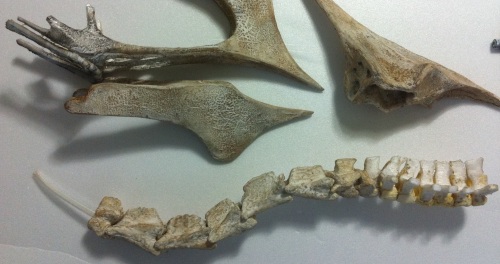
Here is a picture of Tapejara compared to an unknown thalassodromid comparing skull and neck. 2013-07-16 12.45.53

But this post is about the vertebrae that come after the neck: the dorsal vertebrae. The first four to seven of these vertebrae is fused in most adult pterosaurs to support the pectoral girdle and is called the notarium. There is no fossil evidence that this occurred in Tapejaras, but none of the specimens used are considered “full grown adult” either (I’m referring to the ones that were published). Some decisions that affected the dorsals also involved the cervicals, so I might also talk a bit about them here.

I had some trouble with my sources. I used the same ones I had used for the cervicals, but several images published in the article about SMNK PAL 1137 had the wrong scales, so the sizes didn’t match. Not even the reconstructions using the bones from the same specimen matched, and the descriptions confused me more than they helped, so I had to rely on other sources. MN 6588-V has several dorsal vertebra but it’s not really a tapejara, and it also has an incompatible scale: if I match the size of the cervical vertebra in that specimen, all the others become too small.

2013-07-18 13.25.00

So I decided to trust the scale I was originally using (from IMCF 1061) which unfortunately has no dorsal vertebrae, and to estimate the size based on Tupuxuara. The problem is that Tupuxuara’s cervical vertebrae are proportionally shorter, and so is the pelvis, so maybe the Tapejara has a narrower and longer body than the Tupuxuara, or maybe it just has a proportionally longer neck. I don’t know.

I checked other reconstructions (drawings and sculptures), but they seem to either have fewer cervicals, or identical cervicals. I observed that both in Tupuxuara and Tapejara (Iwaki specimens) there are some longer cervicals and others which are shorter and taller. I am not sure about the order, but in the (unpublished) Iwaki specimen, they were numbered, and the two shorter ones were cervicals no. 3 and 5 (not counting Atlas/Axis). I don’t know how accurate that ordering is since there is no publication, but I assume they either knew before preparation or they fit somehow, and since that’s the best information I have, I followed that same order in Tapejara.

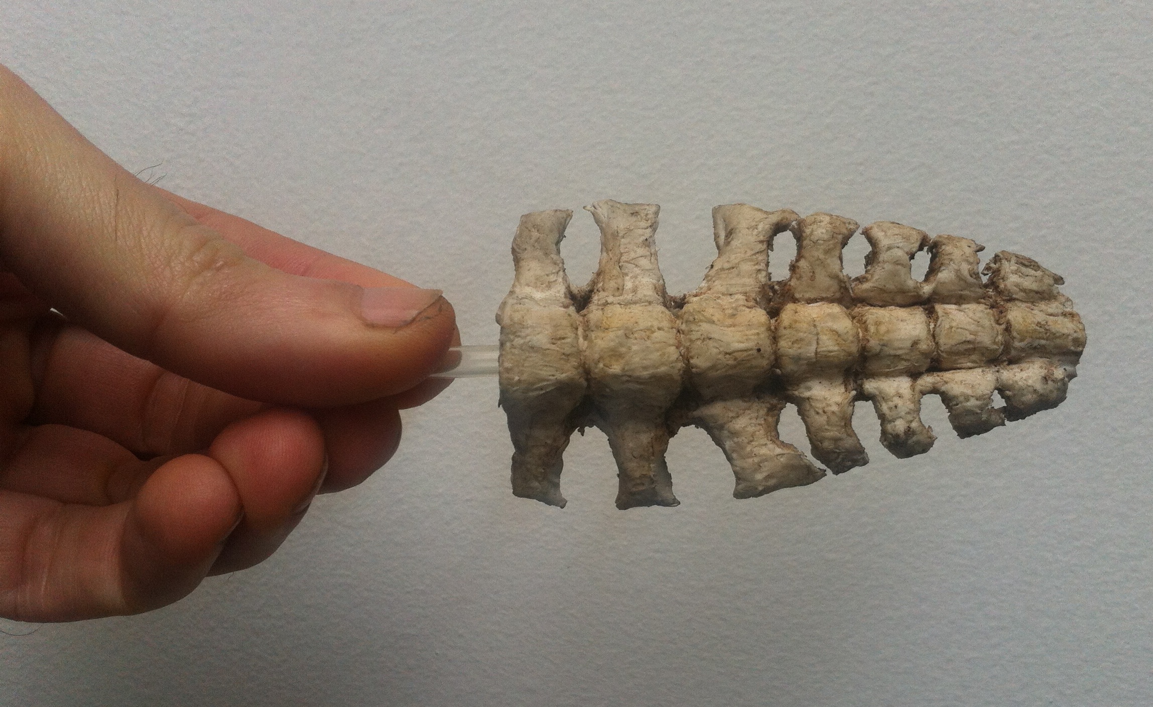
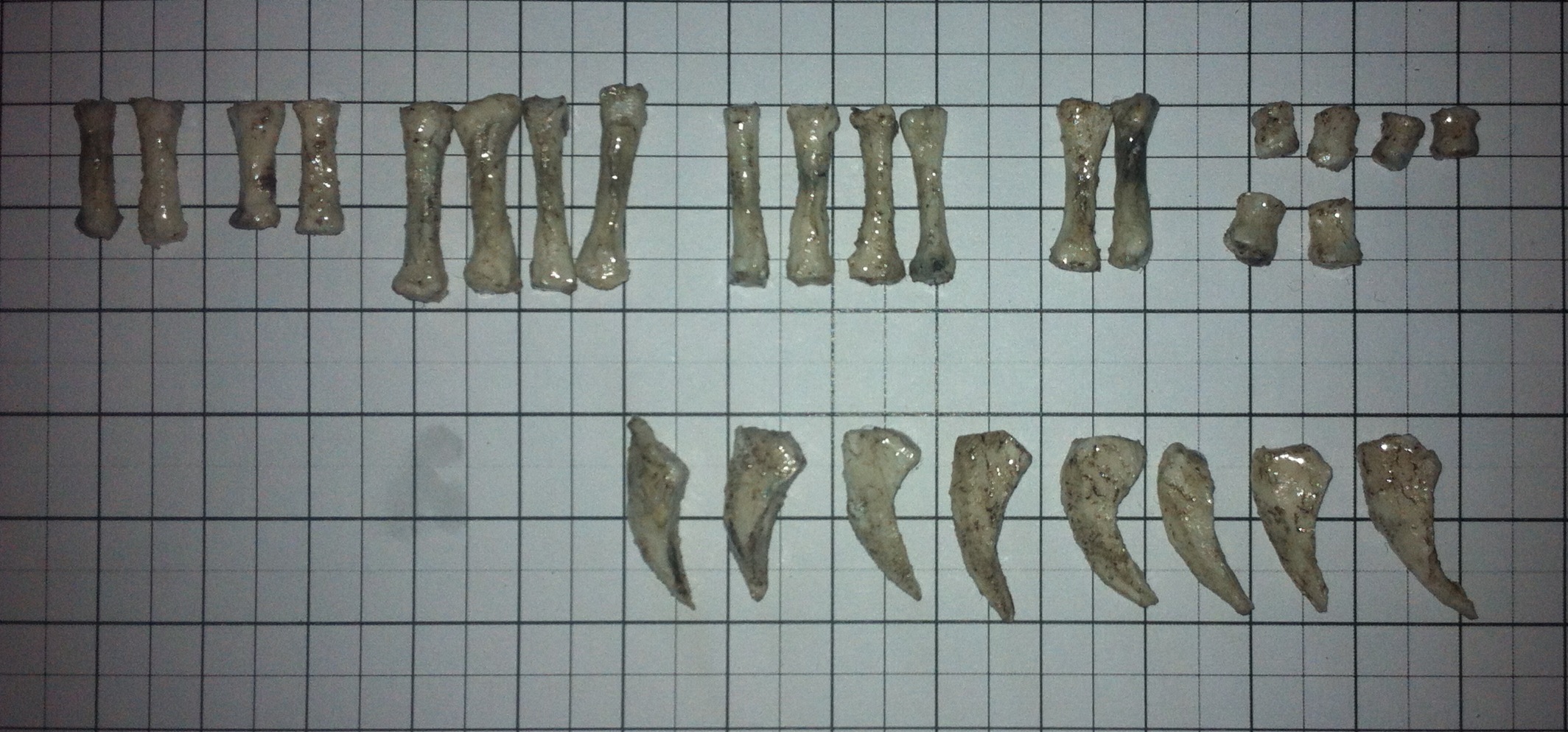
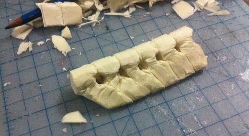
I decided to not use any reconstructions as sources except my own. I used the four dorsal vertebrae from SMNK PAL 1137 as sources ignoring their  (incorrect) scale, adding broken and missing parts, and tried to fit them with the eighth cervical vertebra, using the Tupuxuara notarium as a reference but making each vertebra a little wider. After trying out three different prototypes, I could finally start making a final version.

I used foam strips for the processes and thicker foam for the vertebra’s body.

2013-07-16 17.38.06 2013-07-16 17.43.09 2013-07-16 17.43.18

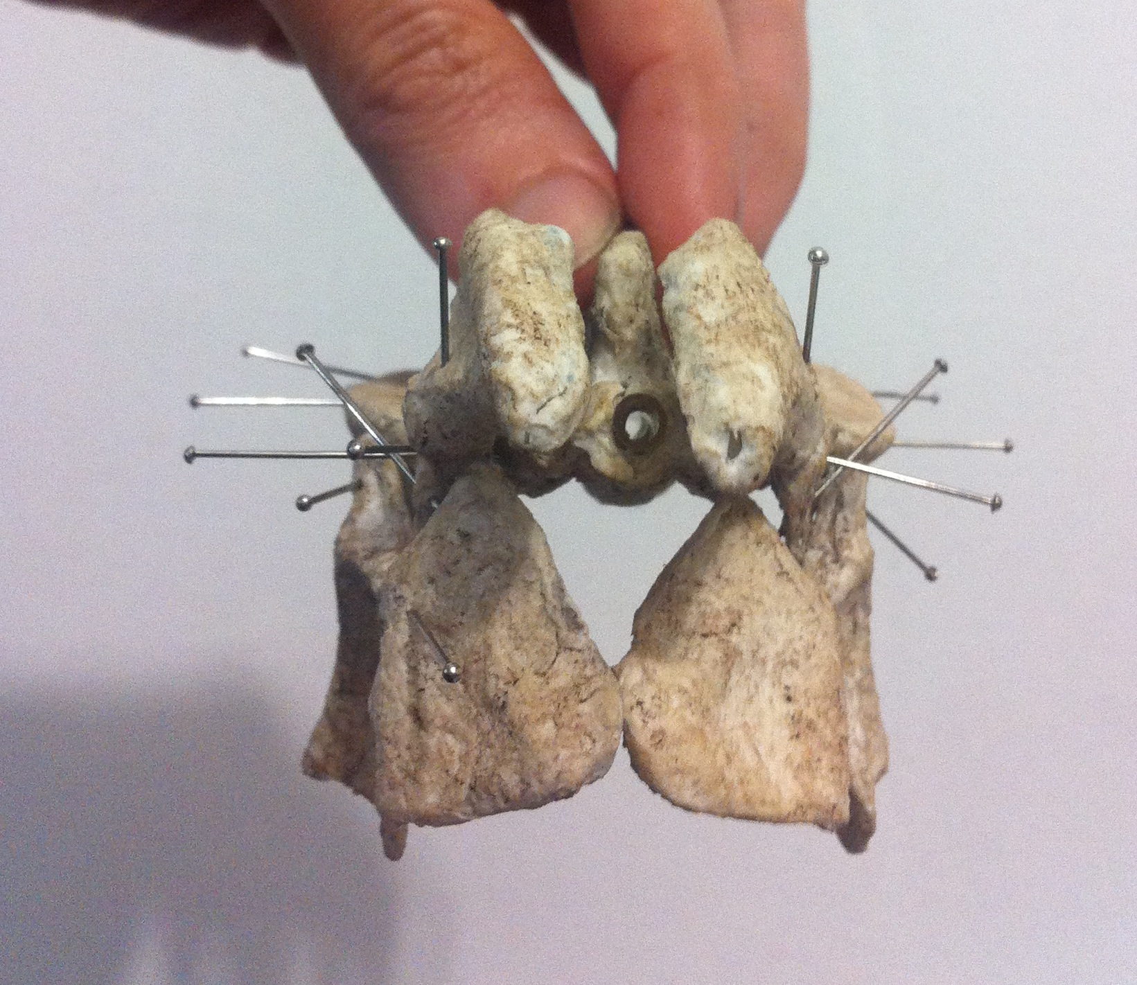
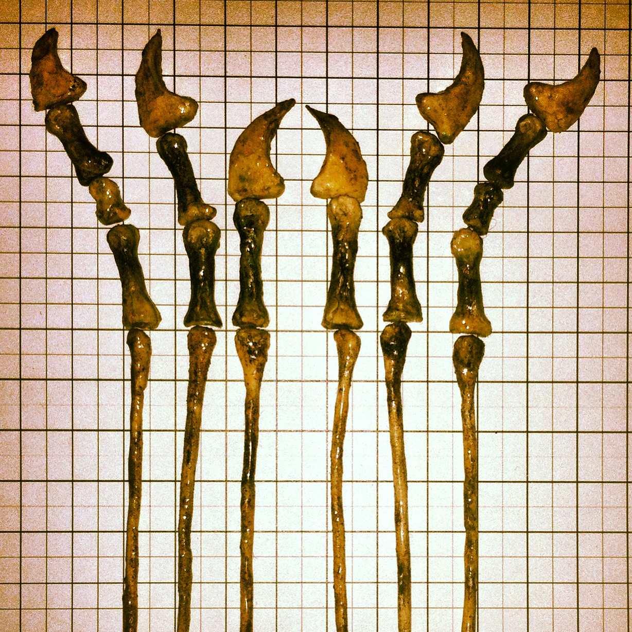
After fitting the processes together, I separated the vertebrae to add details (processes) to each one.

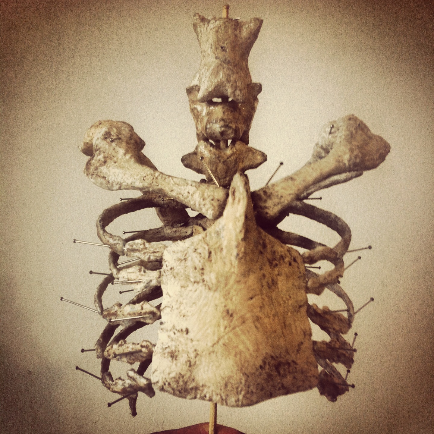
Here are some views of the vertebrae after molding with fire, but before adding modeling resin.

2013-07-16 18.54.44 2013-07-17 16.50.36 2013-07-17 16.50.48 2013-07-17 17.09.58

And here some more views after resin and staining with coffee.

2013-07-18 01.15.54 2013-07-18 13.01.35 2013-07-18 13.02.11 2013-07-18 13.02.28 2013-07-18 13.11.55 2013-07-18 13.20.54

These are the dorsals that are part of the pectoral girdle, and which will articulate with the scapula. The exact number of dorsals after them before the sacrum seems to be unknown. Most reconstructions draw six notarium vertebrae + three to four free ones + two to three pelvic dorsals before the sacrum (that means 5 – 7 more dorsals), but other authors mention 10, 12. It seems that there is not enough evidence to be sure. I am also not sure about their shape. Do they get smaller, narrower, wider?

I don’t mind reading dozens of publications. It’s great. They revealed that many of my assumptions were bad theories, but they usually don’t offer me a solution. They open a discussion. And so, the more I read, the harder it is to make decisions, and I also reach the point where paleontologists disagree. It’s a fantastic discussion, really, but I have a deadline, so I have to make some choices. If they are bad, I will try to fix it before I deliver the skeleton.

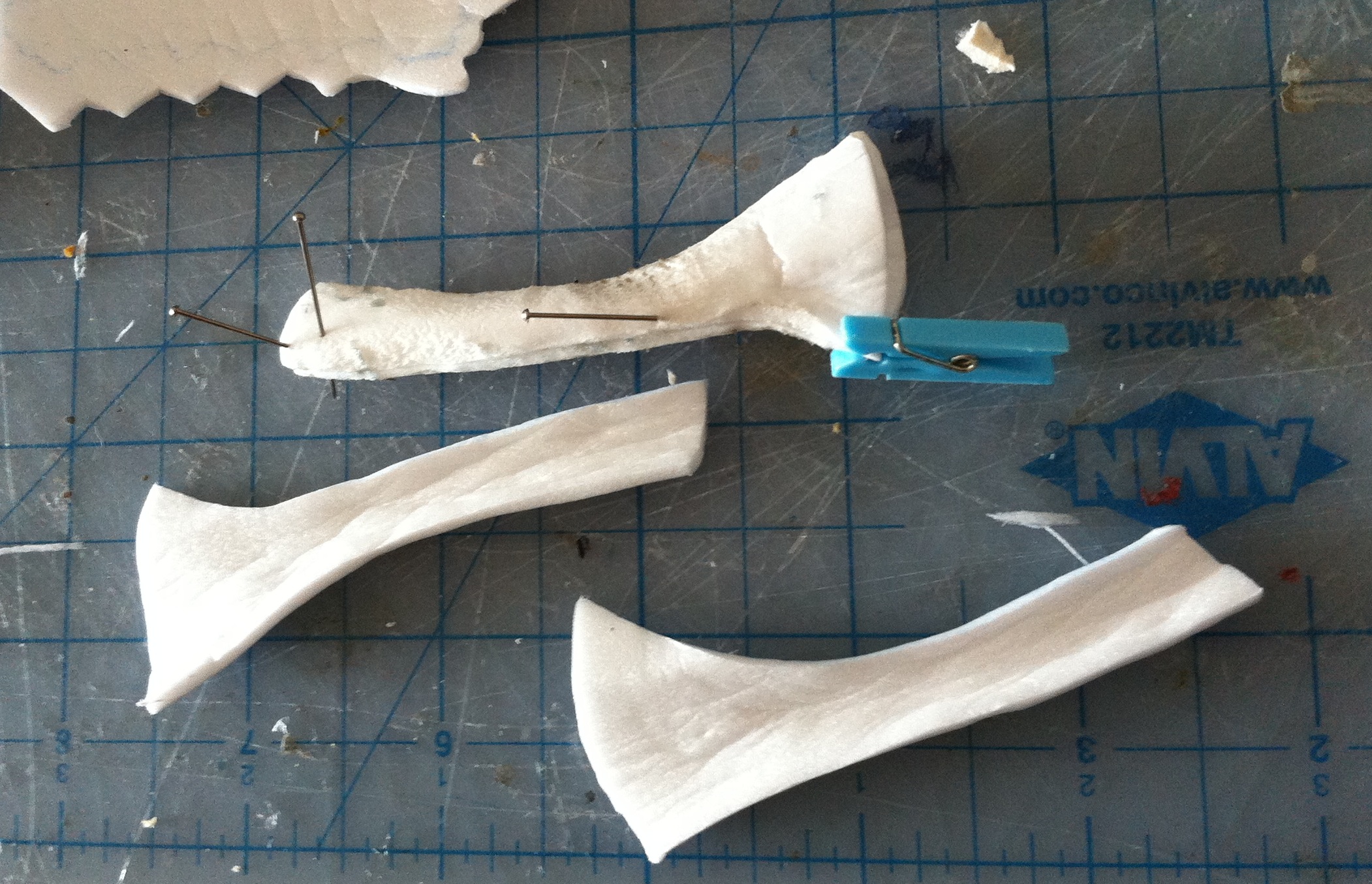
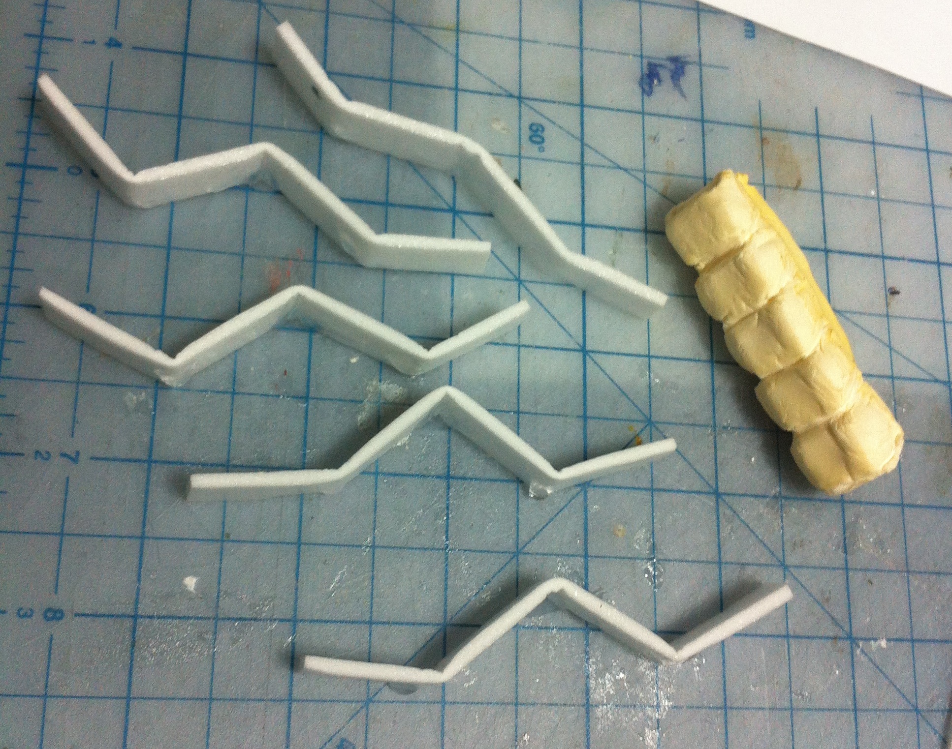
So I decided to make 9 more dorsals (a total of 15). Three will be fused to the pelvic girdle, and the other six will be free. This is a first 3D sketch (the body and the spine of six dorsals).

IMG_5352

After adding the processes and splitting.

IMG_5358

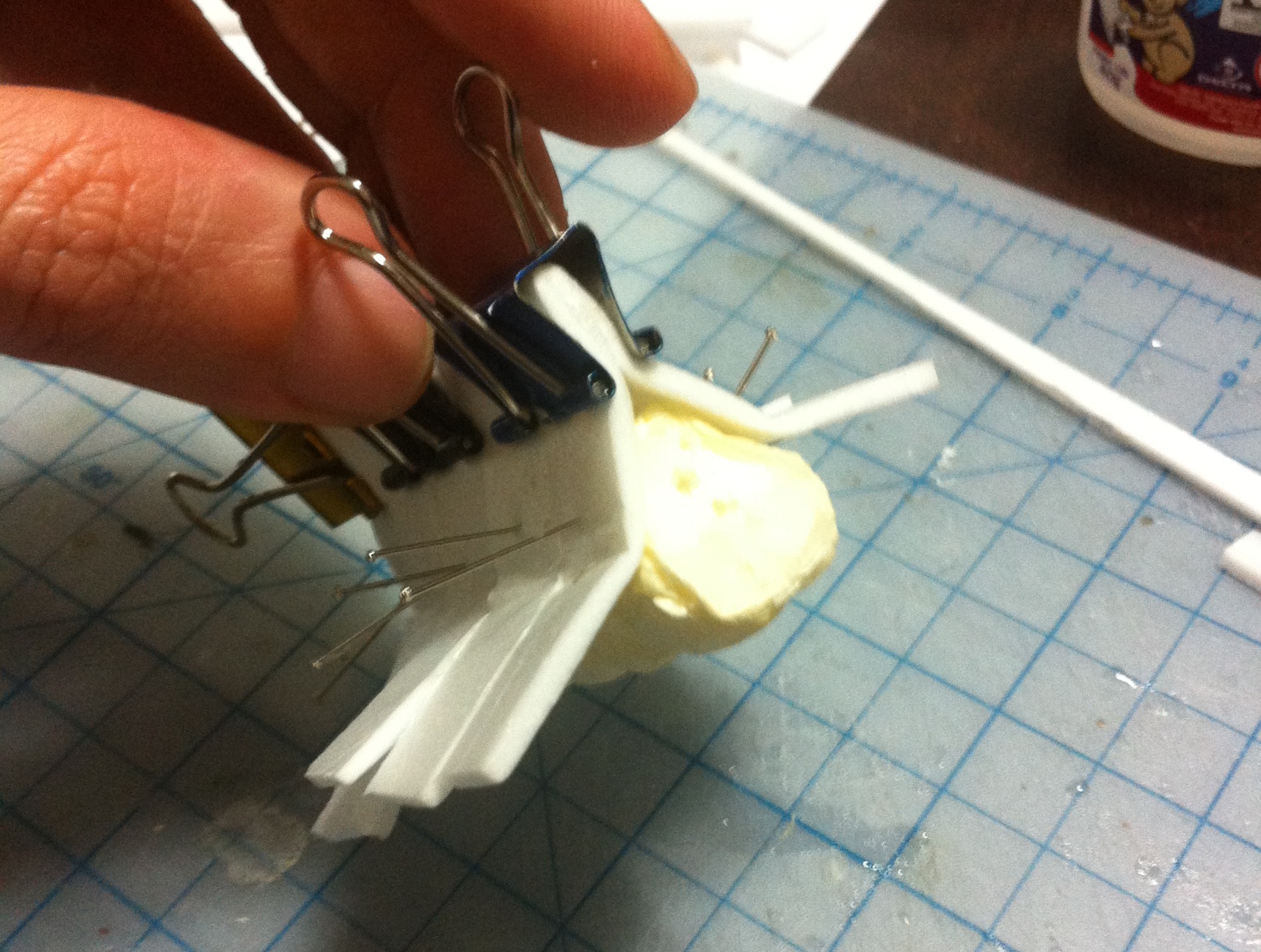
After acrylic resin and staining.

2013-07-25 16.49.18

They are not really finished: I’m using them as prototypes so I can continue the work. Later I might reshape them, cut some excess foam, make them thinner, narrower, or even throw them away and make new ones. For now they just need to be functional.

I’m trying to find reliable information to make the pelvis. The only sources I have are SMNK PAL 1137 (just pelvis, no sacrum, one or two views of each bone and no more), and a side view from MN 6588-V. I would be nice to have other views. I’ve been trying to obtain permission from the Iwaki museum so I can have access to all Tapejara photos (I only have some), but so far I haven’t received any response. I might have to continue without them, unfortunately.

1 Comment

Filed under News and Exhibits, Pterosaur #7: Tapejara